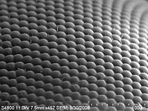
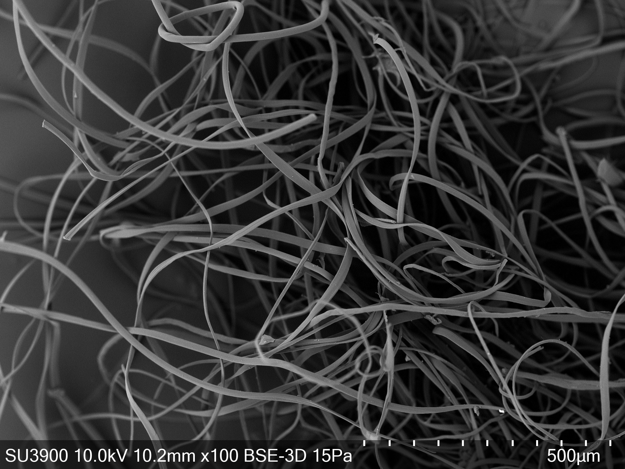
SEM micrographs of filament surfaces at two different magnifications: a

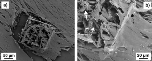
Download scientific diagram | SEM micrographs of filament surfaces at two different magnifications: a , b PCL+1 % CHX; c , d PCL+2 % CHX; e , f PCL+4 % CHX from publication: Combining in the melt physical and biological properties of poly(caprolactone) and chlorhexidine to obtain antimicrobial surgical monofilaments | Bacterial infections on a sutured wound represent a critical problem, and the preparation of suture threads possessing antimicrobial properties is valuable. In this work, poly(caprolactone) (PCL) monofilaments were compounded at the concentration of 1, 2 and 4 % (w/w), | Chlorhexidine, Antimicrobials and Antiseptics | ResearchGate, the professional network for scientists.
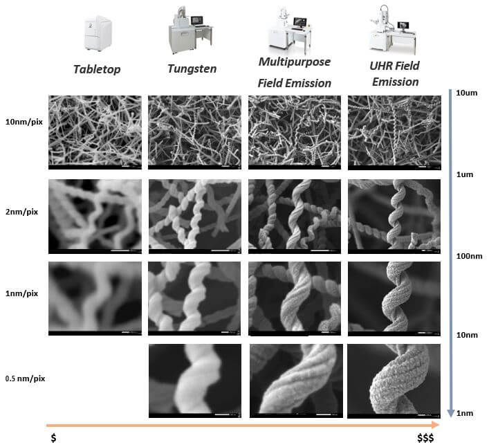
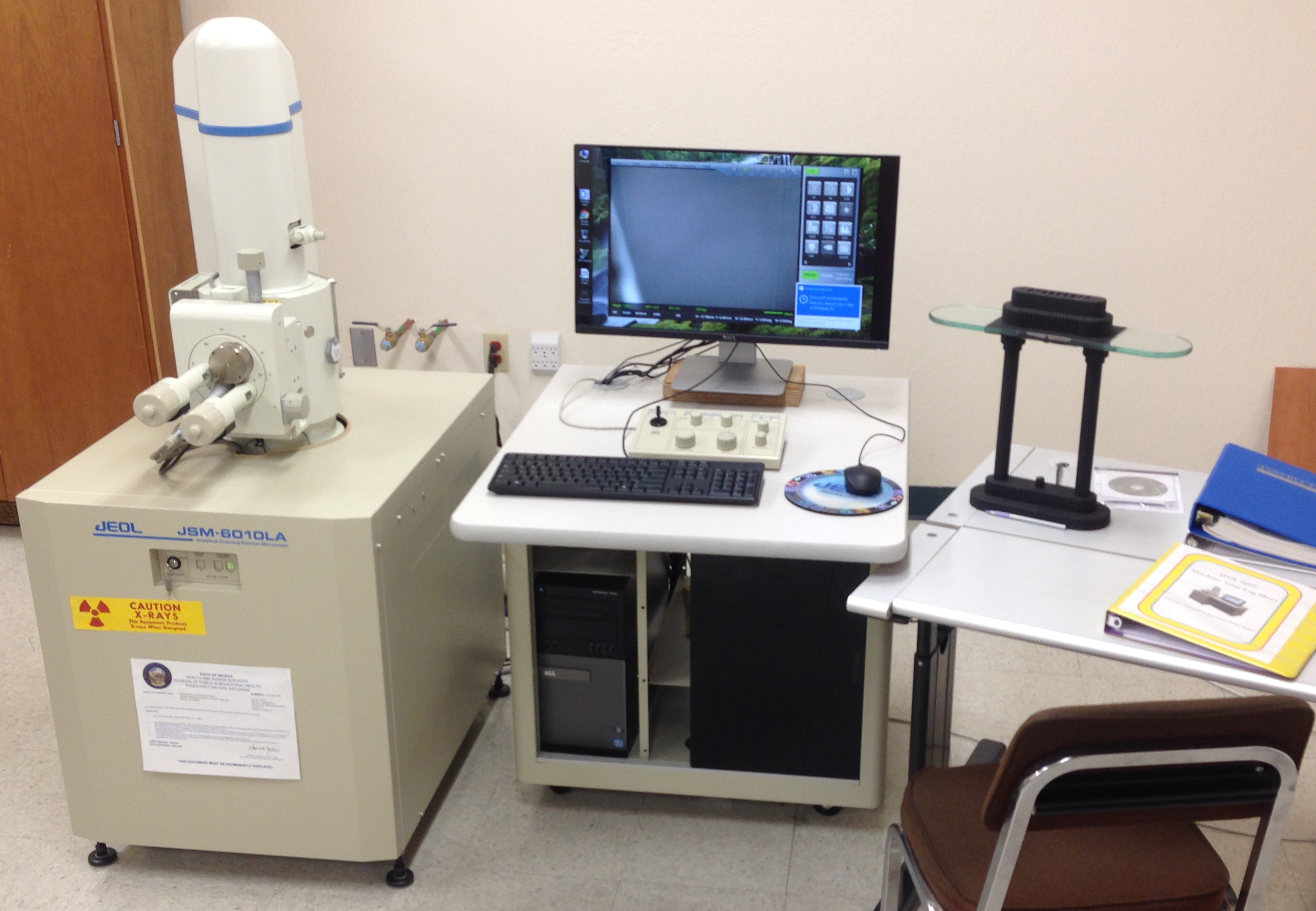
JEOL USA blog Choosing the right scanning electron microscope f

Scanning Electron Microscopy
Scanning electron microscope - Wikipedia
Mackay School of Earth Science and Engineering Microbeam Laboratory
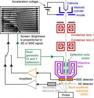
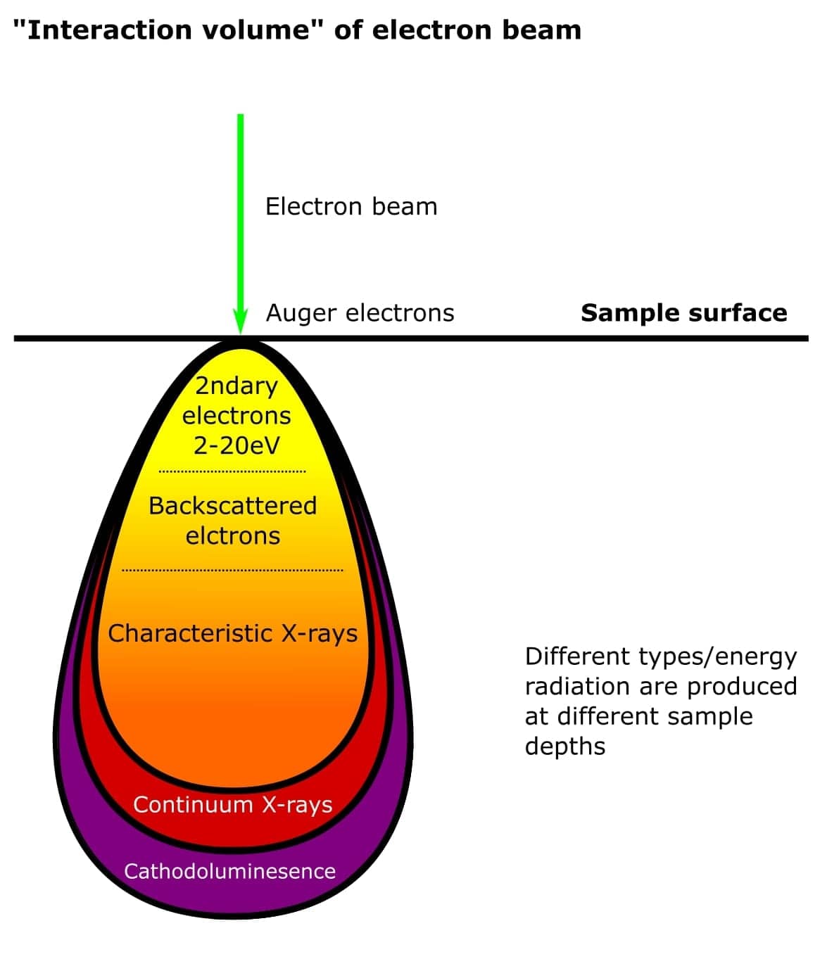
SEM Principle West Campus Materials Characterization Core
What Is An Electron Microscope? 4 Types Of EM - VacCoat
Scanning Electron Microscopy (SEM) - Surface Science Western
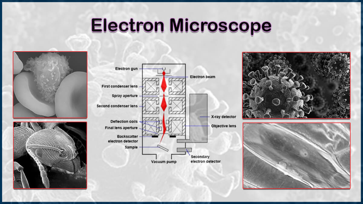
Types of Electron Microscopes
The 2 Main Electron Microscopy Techniques Explained

A cool guide to what goes on inside a scanning electron microscope

Dr nh mat.char notes
EE.1943-7870.0001502/asset/18ff4d28-43d8-4bd3-a9ac-b5dd32b0237e/assets/images/large/figure3.jpg)
Potential of a Static Magnetic Field to Inhibit Filamentous Sludge
High Resolution Cryo Scanning Electron Microscopy of
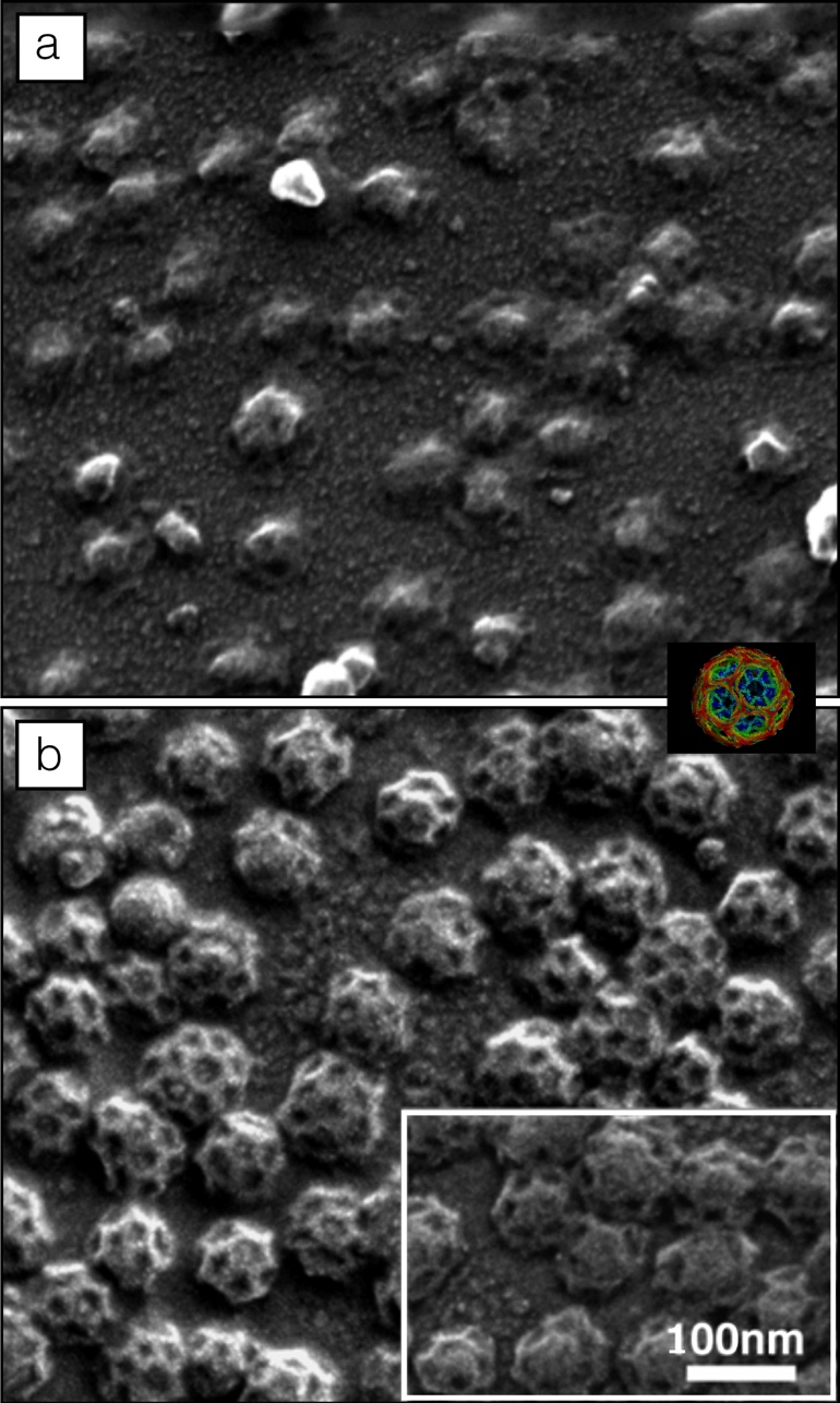
SEM images at two different magnifications of a cage mi