BB700 Mouse Anti-Rat RP-1 Antigen
lt;span style="font-family:Times,serif;color:#000000;font-size:9pt;">The RP-1 monoclonal antibody specifically recognizes the RP-1 Antigen. This cell surface marker is expressed on rat peritoneal and peripheral blood neutrophils. Amongst bone marrow cells, the RP-1 Antigen is expressed on band form and mature neutrophils but is not expressed on promyelocytes, myelocytes, and metamyelocytes. The RP-1 antibody does not bind to either rat monocytes, macrophages, eosinophils or to peritoneal neutrophils from mice, rabbits, guinea pigs, or to human peripheral blood neutrophils. Expression of the RP-1 Antigen on rat peritoneal neutrophils is enhanced by cellular stimulation with Phorbol 12-Myristate 13-Acetate (PMA) or Concanavalin A (ConA). Immunoprecipitation and SDS-PAGE analysis of non-treated and PMA-activated rat neutrophil membranes with the RP-1 antibody revealed two main bands of approximately 85 kDa. The RP-1 antibody is also known as the Mouse Anti-Rat Granulocytes antibody.</span>
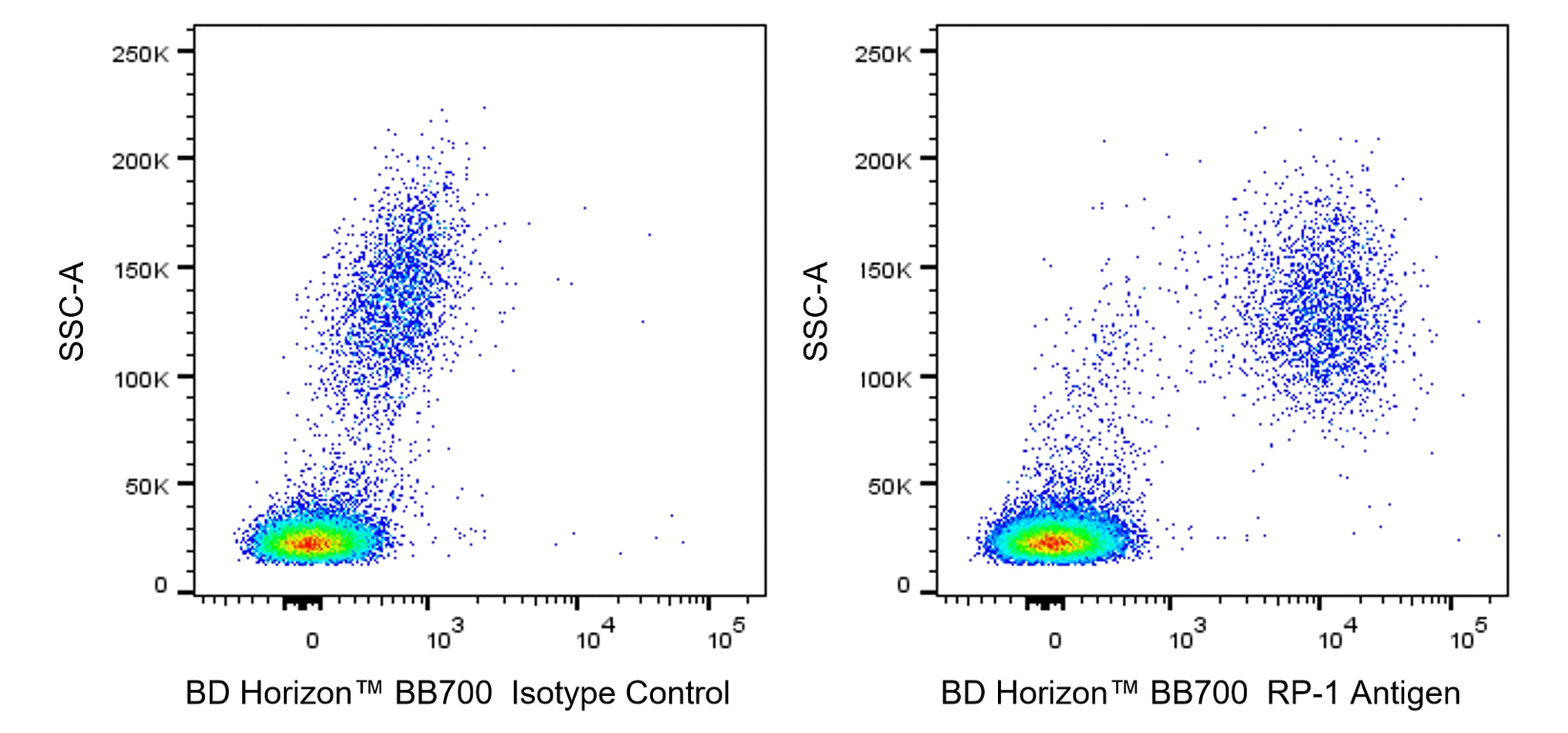
Multiparameter flow cytometric analysis of RP-1 Antigen expression on rat peripheral blood leucocytes. Rat whole blood was treated with BD Pharm Lyse™ Lysing Buffer (Cat. No. 555899) to lyse erythrocytes. The leucocytes were washed and stained with either BD Horizon™ BB700 Mouse IgG2a, κ Isotype Control (Cat. No. 566419; Left Plot) or BD Horizon BB700 Mouse Anti-Rat RP-1 Antigen antibody (Cat. No. 566871; Right Plot) at 1 µg/test. A two-parameter pseudocolor density plot showing the correlated expression of RP-1 Antigen (or Ig Isotype control staining) versus side-light scatter (SSC-A) signals was derived from gated events with the forward and side-light scatter characteristics of viable leucocyte populations. Flow cytometry and data analysis were performed using a BD LSRFortessa™ Cell Analyzer System and FlowJo™ software. Data shown on this Technical Data Sheet are not lot specific.

BB700 Rat Anti-Mouse CD162
Mouse Monoclonal Antibody

RP-1 Antigen Mouse anti-Rat , BB700, Clone: RP-1, BD Biosciences™

Feeding mode influences dynamic gut microbiota signatures and
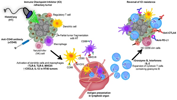
Boiling histotripsy and in-situ CD40 stimulation improve the
BB700 Mouse Anti-Rat RP-1 Antigen
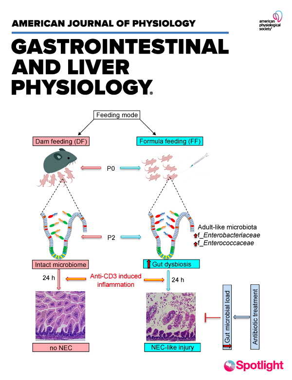
Feeding mode influences dynamic gut microbiota signatures and
Anti-Iba1 antibody [HL22-RT] (GTX635400)

Feeding mode influences dynamic gut microbiota signatures and

Flow Cytometry
Mouse Monoclonal Antibody

RP-1 Antigen Mouse anti-Rat , BB700, Clone: RP-1, BD Biosciences™